

Research
We study how hosts and diseases interact inside intact tissue. We build the technologies this needs, mostly for high-dimensional tissue imaging, and use existing ones where they fit.
We ask how the immune system responds to viruses such as HIV, SIV, SARS-CoV-2, EBV and Ebola virus, to cancers such as lymphoma, and in immune diseases such as chronic rhinosinusitis. With clinical collaborators, we work from archival clinical samples, small and large animal models and organoids. The long-term goal is better therapies.
Technology development
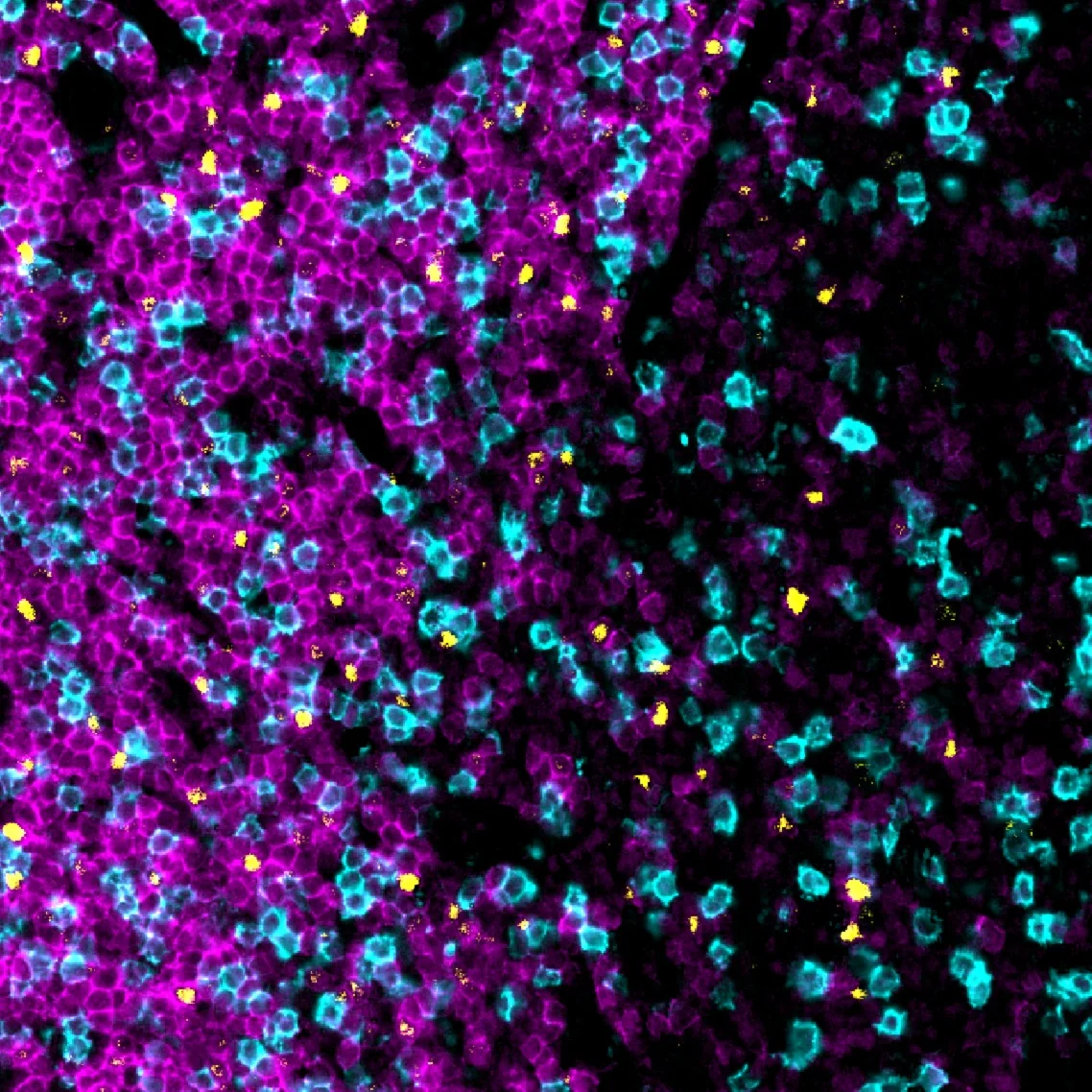
We develop chemistry and imaging that measure proteins, RNA, DNA and glycans in the same tissue section.
- PASTA
- Tyramide amplification of oligonucleotide barcodes, for high-plex protein and RNA imaging on the same FFPE section.
- IN-DEPTH
- Single-cell protein imaging guides RNA capture on the same slide, without losing RNA signal. Our SGCC framework then links the two.
- PANINI
- Protein and nucleic acid imaging in the same archival tissue, sensitive down to single genomic events.
- ExPRESSO
- Tissue expansion that holds under vacuum, for sharper high-plex images on MIBI and imaging mass cytometry.
- GlycoScope
- Human lectins read out glycans together with RNA, in single cells and in tissue.
- MicroCart
- Maps host cells and gut microbes together, across protein, RNA and glycan imaging.
- EpicMIBI
- Imageable epitope barcodes that track tumor clones inside tissue.
- REDSEA
- Corrects signal that spills between neighboring cells in multiplexed images.
- MIBI guide
- Our MIBI workflow for FFPE tissue, step by step.
AI for biology and medicine
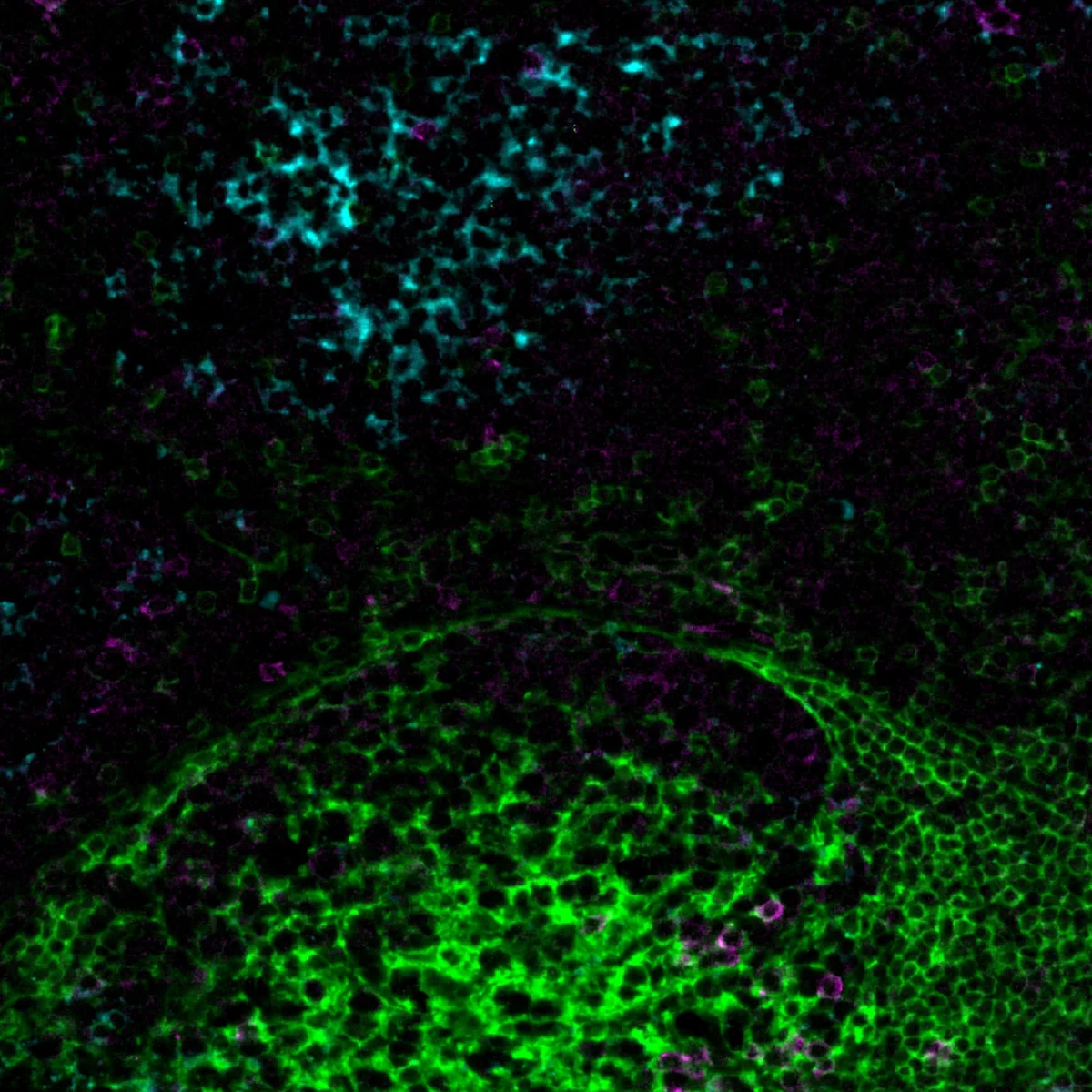
We build models that learn from single cells, their neighborhoods and whole tissue images.
- KRONOS
- A foundation model for spatial proteomics, trained on more than 47 million image patches covering 175 protein markers.
- CellLENS
- Learns cell identities from markers, neighbors and tissue images together.
- MAPS
- Pathologist-level cell type annotation from spatial proteomics images.
- MARIO
- Matches single cells across datasets that share only a few markers.
- COSIE
- Combines histology, epigenome, transcriptome, proteome and metabolome into one map, across tissue sections, individuals and platforms.
Human-centered discovery and translation
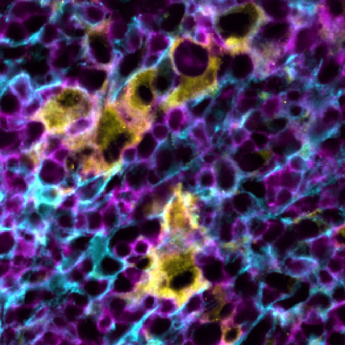
We study viral infection, cancer and immune disease in human tissue. With our clinical partners, we work to turn what we find into better care for patients.
Viral pathogenesis
Viruses are the best molecular biologists. We look at virus-host interactions in their native tissue, to see how viruses get around host defenses. Projects include HIV, SIV, SARS-CoV-2, EBV and Ebola virus.
Selected papers
- A Human Genome-wide CRISPR-Cas9 Screen Reveals Host Factors That Support Epstein-Barr Virus B cell infection
- Same-Slide Spatial Multi-Omics Integration with IN-DEPTH Reveals Tumor Virus-Linked Spatial Reorganization of the Tumor Microenvironment
- Coordinated immune-epithelial dynamics in the nasal epithelium protect against respiratory virus infection
- Epstein-Barr virus orchestrates spatial reorganization and immunomodulation in the classic Hodgkin lymphoma tumor microenvironment
- Epstein-Barr virus exploits desmocollin 2 as the principal epithelial cell entry receptor
- A DNA tumor virus globally reprograms host 3D genome architecture to achieve immortal growth
- Combined protein and nucleic acid imaging reveals virus-dependent B cell and macrophage immunosuppression of tissue microenvironments
- Rhesus Macaque CODEX Multiplexed Immunohistochemistry Panel for Studying Immune Responses During Ebola Infection
- Determinants of SARS-CoV-2 entry and replication in airway mucosal tissue and susceptibility in smokers
- ACE2 localizes to the respiratory cilia and is not increased by ACE inhibitors or ARBs
- The Epstein-Barr Virus Regulome in Lymphoblastoid Cells
- Epstein-Barr virus oncoprotein super-enhancers control B cell growth
- Epstein-Barr virus nuclear antigen 3A partially coincides with EBNA3C genome-wide and is tethered to DNA through BATF complexes
- Epstein-Barr virus nuclear antigen 3C binds to BATF/IRF4 or SPI1/IRF4 composite sites and recruits Sin3A to repress CDKN2A
Cancer biology
“Cancer cells grow faster, adapt better. They are more perfect versions of ourselves” (Siddhartha Mukherjee, The Emperor of All Maladies). We study cancers in their native tissue, to find better ways to predict and treat them. Projects include lymphomas, leukemia and pancreatic cancer.
Selected papers
- Spatial Proteomics Uncovers Immune Cell Neighborhoods and Tumor-Immune Interfaces in Pancreatic Ductal Adenocarcinoma
- Follicular Lymphoma Transformation is Characterized by Cytokine-associated Remodeling of Stromal and Macrophage Compartments
- Same-Slide Spatial Multi-Omics Integration with IN-DEPTH Reveals Tumor Virus-Linked Spatial Reorganization of the Tumor Microenvironment
- Epstein-Barr virus orchestrates spatial reorganization and immunomodulation in the classic Hodgkin lymphoma tumor microenvironment
- Diffuse Large B-cell Lymphomas Have Spatially-Defined Tumor-Immune Microenvironments Revealed by High-Parameter Imaging
- A DNA tumor virus globally reprograms host 3D genome architecture to achieve immortal growth
- Spatial epitope barcoding reveals clonal tumor patch behaviors
- Highly Multiplexed Phenotyping of Immunoregulatory Proteins in the Tumor Microenvironment by CODEX Tissue Imaging
- Multimodal Analysis of Composition and Spatial Architecture in Human Squamous Cell Carcinoma
- Epstein-Barr virus oncoprotein super-enhancers control B cell growth
Immune dysregulation
How do our defenders become the enemy? We study how the body’s defenses go awry, to build better tools to keep them in check. Projects include HIV, lymphomas, chronic rhinosinusitis, inflammatory bowel disease, autoimmune hepatitis and neural-immune dysfunction.
Selected papers
- Coordinated immune-epithelial dynamics in the nasal epithelium protect against respiratory virus infection
- Epstein-Barr virus orchestrates spatial reorganization and immunomodulation in the classic Hodgkin lymphoma tumor microenvironment
- Multi-scaled Transcriptomics of Chronically Inflamed Nasal Epithelium reveals Immune-Epithelial Dynamics and Tissue Remodeling in Nasal Polyp Formation
- A multi-omics spatial framework for host-microbiome dissection within the intestinal tissue microenvironment
- Combined protein and nucleic acid imaging reveals virus-dependent B cell and macrophage immunosuppression of tissue microenvironments
- Inflammatory molecular endotypes of nasal polyps derived from White and Japanese populations
Join us
We are always looking for talented future colleagues on both the computational and experimental sides.